Central Research Institute of Skin and Venereal Diseases of the Federal Agency for Health and Social Development, Moscow.
The contour plastic technique is a popular method for the correction of involutional changes in the skin. Collagen and hyaluronic acid are most frequently used for the purpose. The reported clinical observations indicate that collagen preparations can be used not only as substitution therapy but also to revitalize tissues and stimulate neocollagenogencsis in the derma.
Aging is a complex biological process characterized by metabolic, structural and functional changes in the cellular structures of tissues. Unlike internal organs, where aging occurs quite latent, signs of this process on the skin are always visible (1).
External signs of skin aging are expressed in the appearance of wrinkles, decreased turgor, dryness, thinning and the appearance of pigmentation. Age-related changes in the skin occur due to numerous exogenous and endogenous causes [2, 3]. Exogenous factors include climatic, chemical, biological and physical factors, i.e. those that are directed at the skin from the outside. Among the physical effects, the influence of UV radiation should be particularly noted, which is why the term “skin photoaging” appeared (2, 3).
Endogenous factors are the cause of natural aging and include a genetically determined program of aging and cell death [4, 5]:
— accumulation of harmful substances by cells and the intercellular matrix during life [6-8];
— changes in hormonal status (9-13);
— decreased immunity [14];
— psycho-emotional stress, etc. [15—17].
Pathomorphological aging of the skin is characterized by a decrease in its thickness, a decrease in the number of fibroblasts, their size, proliferation rate, and smoothing of the dermalepidermal border. A decrease in the synthetic activity of dermal cells leads to a decrease in their production of such important components of the main substance of the dermis as dermatan sulfate, chondroitin sulfate and hyaluronic acid. Studies show that the amount of hyaluronic acid in aging skin decreases [11], while the amount of chondroitin sulfate, on the contrary, increases [18].
A decrease in the amount of hyaluronic acid leads to a decrease in hydration, turgor and elasticity of the skin, contributing to dryness and the formation of wrinkles. In aging fibroblasts, the production of tissue inhibitors of metalloproteinases also decreases, while the synthesis of collagenases, gelatinases, and elastase increases [19]. Collagen content accounts for 70 % of the dry mass of the dermis, with type 1 collagen accounting for 80 % and type 3 collagen accounting for approximately 15 % of the total collagen volume. Throughout a person’s life, collagen content decreases by approximately 1% per year, with the ratio of collagen types changing towards an increase in the content of type 3 collagen, primarily due to a decrease in the content of type 1 collagen [20, 21]. Despite a significant reduction in the number of collagen fibers, their network in old skin becomes even denser than in young skin. Most collagen fibers lose their former ability to stretch, thicken and become woven into bundles with a random orientation, unlike young skin, where they are in an ordered state. Elastin, which makes up only 2 % of the total volume of proteins in the dermis, along with collagen fibers, will give it firmness and elasticity. By the age of 70, degenerative changes are observed in most elastic fibers; In this case, characteristic cysts and lacunae are formed in the reticular layer of the dermis, and a network of thin elastic fibers is formed within the papillary layer of the dermis. The dermis of old people is relatively poorly vascularized. Compared with the skin of young people, the skin of old people has significantly fewer vertical capillary loops in the papillary dermis. In addition, a sharp (35 %) decrease in the number of venules is detected, which is probably associated with a 50% decrease in the number of tissue basophils involved in the synthesis of heparin, a powerful angiogenic factor [20].
Decreased vascularization leads to a deterioration in the supply of nutrients to the skin, reduces thermoregulation, lowering the surface temperature of the skin.
The aging process does not bypass the skin appendages: the number of eccrine sweat glands decreases and their function declines, the rate of nail growth decreases by 40-50 %, the number, diameter of hair and the rate of its growth decrease. Gray hair, one of the manifestations of aging, is a consequence of a decrease in the melanin-forming function of melanocytes in the hair follicles, on the one hand, and of a genetic predisposition, on the other hand. Paradoxically, the sebaceous glands, on the contrary, begin to increase in size with age, but the production of fat in them still decreases. Localized increase in the number of melanocytes in small areas of irradiated skin and simultaneous disruption of melanosome transport to keratinocytes lead to mottled pigmentation, a marker of photoaging.
Immunohistochemical studies within the papillary layer of the dermis exposed to insolation reveal a 20-30 % decrease in the content of collagen types 1 and 3, which may be a consequence of increased breakdown, on the one hand, and of a decrease in its synthesis by fibroblasts, on the other hand (18, 211). In addition, in the dermis, which is more susceptible to photoaging than to chrono aging, the ratio of the concentration of collagen types 3 and 1 also changes: the amount of the latter decreases by 59 %, and this decrease correlates with the severity of photodamage to the skin |22, 23|. Thus, aging skin, regardless of the form of aging, is characterized by changes in the structure of the collagen protein.
Collagen (Greek: kolla — glue, genes — giving birth) is a fibrillar secretory protein, the most common in the human body. It is a component of all tissues that require a framework or mechanical support to give them their characteristic shape. Depending on the composition and percentage of amino acids, different types of collagen are distinguished. Type 1 collagen is the most widely represented; it makes up 90 % of all collagen in the body and is found in skin, bones, cartilage, tendons and other tissues.
Collagen forms fibers that intertwine like threads in tissue and creates a framework into which new cells can grow. In this way, collagen in human skin provides its elasticity and firmness, and disruption of its synthesis leads to the loss of these properties.
Currently, more than 20 genetically distinct types of collagen have been identified. These molecules consist of 3 polypeptide chains of different types (α-helices), twisted into a triple righthanded helix. In turn, polypeptide chains are constructed from frequently repeating fragments with the characteristic sequence GLY-X-Y.
Every third amino acid residue is glycine, the X position is occupied by proline, and the Y position is occupied by both proline and hydroxyproline. The presence of hydroxyamino acid residues in the polypeptide chain is a characteristic feature of collagen. This determines its rigidity.
The role of these amino acids is extremely important in stabilizing the triple-helix conformation of the collagen molecule. At one end, the collagen molecule is cross-linked, the number of which increases as the body ages.
Based on morphology, collagen is usually divided into three groups:
— fibrillar collagen: type 1, 2, 3, 5 and 11 collagens;
— reticular collagen —type 4 collagen that forms the supporting network of basement membranes;
— filamentous collagen — molecules of collagen type 6.
In the adult human dermis, interstitial fibrillar collagen (types 1, 3, and 5) is the largest fraction of collagen: approximately 80-90 % is type 1 collagen and 8-12 % is type 3 collagen. Correction of age-related changes is one of the most pressing issues. The prerogative is given to methods with a minimal rehabilitation period.
Taking into account the above-mentioned changes occurring in the dermis with age, a wide range of products is used for their therapeutic correction. The most widely used preparations are hyaluronic acid and collagen.
In world practice, collagen is the most popular, widely used and promising biomaterial. Provided that the three-helix structure of the fiber is preserved, it is able to grow through these fibers into the adjacent tissue, stimulating it to renew and grow, and thus acts as a control matrix. Subsequently, it is absorbed and utilized, and the
products of lysis participate in metabolic reparation processes. For over 30 years, type 1 collagen preparations obtained from cattle skin have been widely used in medicine. The collagen is closest to human collagen in its biological composition and structure. However, the effect of these products was not optimal due to the lack of strong covalent intermolecular bonds and disruption of the fiber integrity. The collagen biomaterial COLLOST® preserves the fibrillar structure, which ensures the regeneration of damaged tissues.
Various forms of COLLOST® are made from cattle (bull) skin based on type 1 collagen, which is as close as possible to human collagen in structure and composition. The method of obtaining this preparation allows preserving the singe structure of the fiber by moving away from the usual accepted method of lyophilization (i.e. thermal treatment).
The new production method will provide a high degree of purification from ballast substances and ensure obtaining of a product that is ideally close to human collagen. COLLOST® gel occupies a special place. The properties of COLLOST® make it promising as a gel for use in cosmetology for wrinkle correction. Along with the correction of cosmetic defects, the production of one’s own autologous collagen is stimulated, which is of particular interest when working with this product.
When using the product to correct age-related skin changes, one should take into account contraindications (infection of the injection area, systemic collagen diseases, allergic reactions to bovine collagen) and possible complications (wound infection, pain at the injection site, allergic rashes, blood stains, bruises at the injection site, swelling). Before administering the product, it is necessary to conduct allergy tests and collect an allergy history.
We present clinical cases of injection of COLLOST® gel in the medical cosmetology department of the Federal State Institution Central Scientific Research Institute of Dermatovenerology of the Russian Ministry of Healthcare. The aim of this study was to investigate the processes occurring in the dermis after the introduction of the collagencontaining product COLLOST®, using the histological method and the ultrasound scanning method. The study involved female patients aged 30 to 65 years. Agerelated changes are represented by a decrease in tissue turgor, deformation of the facial oval, nasolabial folds, the presence of static and expression wrinkles (“crow’s feet”, transverse forehead wrinkles, wrinkles between the eyebrows), dullness of complexion, and thinning of the skin.
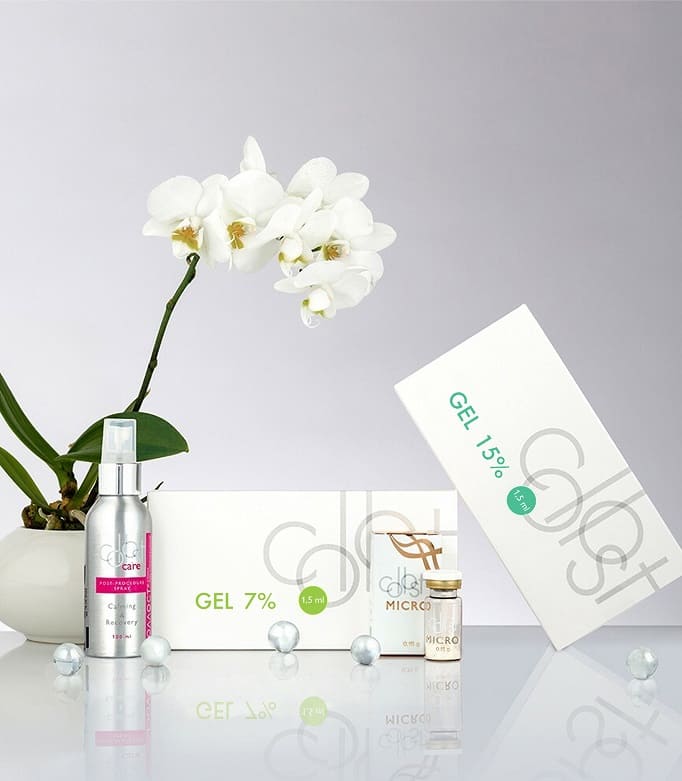
The clinical effect of the therapy was assessed using photography before and after the injection of COLLOST®. To objectify the obtained data, a histological examination and ultrasound scanning of the skin were carried out before, during and after the course of treatment. For administration, the product COLLOST® was used in sterile ampoule- syringes 7 and 15% for contour plastic surgery in the correction of nasolabial folds and fine wrinkles.
The product was injected intradermally using the standard accepted method. The method of introduction is tunnel. The frequency of injection is three times, with intervals of 2 and 4 weeks after the first injection of the product. COLLOST® gel has a high viscosity, which makes it difficult to administer intradermally at room temperature. Before injection, the product was warmed up to human body temperature. Correction was performed on wrinkles in the nasolabial fold area, wrinkles at the corners of the mouth, “crow’s feet”, transverse forehead wrinkles, and wrinkles between the eyebrows.
The product was administered in a volume of 1–1.5 ampoules into each correction area. Moderate swelling that occurred after the product was administered persisted for an average of 1–3 days and resolved on its own.
After the course of injections, an improvement in turgor, color, face contour and skin texture was observed in the areas that underwent correction. The elimination of static wrinkles reached 80 % of the original (see Fig. 1 and 2 in the color insert). Histological examination was performed three times: one day after the initial administration of the product, 14 days before its repeated administration, and 14 days after the repeated administration of the product.
During the initial histological examination, cavities filled with homogeneous protein masses that did not have a fibrillar structure were visible in the middle part of the dermis (see Fig. 3a in the color insert); there was no inflammatory reaction, and the structure of the epidermis was not damaged.
During a repeated histological examination after 14 days, the skin had a normal structure, the dermis was represented by collagen and elastic fibers of normal structure, and no inflammatory reaction or changes in the structure of the epidermis were detected. Histological examination after double administration of the product showed a significant increase in collagenogenesis. In the middle part of the dermis, in the area of the product injection, basophilic protein masses are visible that do not have a fibrillar structure, with a pronounced macrophage reaction around them. In the peripheral areas, an increase in the number of fibroblasts and an increase in the formation of collagen fibers were noted, which were represented by thin basophilic fibrils forming a network between the mature fibrillar structures of the dermis (see Fig. 3b in the color insert).
Ultrasound scanning data indicate an increase in the echogenicity of the dermis, an increase in its thickness, and an increase in neoangiogenesis, which allows us to speak about biorevitalization of tissues in the correction zones (see Fig. 4 on the color insert). An increase in the number of small vessels in the dermis was also noticeable. There were no changes in the epidermis. Thus, double injection of the product leads to the development of a macrophage reaction in the injection area, which indirectly activates fibroblasts and increases the synthesis of collagen fibers in the dermis.
Thus, the use of the product COLLOST® is promising as a bioimplant for the correction of age-related skin changes.

Fig. 1. Clinical effect of COLLOST® gel injections into the nasolabial fold area.
a — before injection of the product; b — 14 days after injection; in — after three-fold injection.

Fig. 2. Before (a) and after (b) injection of COLLOST® gel 15 % into the nasolabial fold area.
A decrease in the depth of nasolabial folds was noted.

Fig. 3. Histological preparation:
a — on the 1st day after injection of COLLOST® gel; b — after double injection (28th day) of the product.

Fig. 4. Ultrasound skin scan data.
a — healthy skin; b — after three-fold injection of COLLOST® gel

Write to us and our specialists respond as soon as possible